|
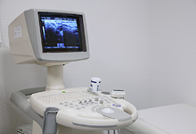
Examination using ultrasound (sonography) is a completely harmless procedure that does not involve any radiation and can even be used without any risks during pregnancy. The principle of sonography is based on the application of ultrasound waves in the inaudible range. The examining doctor receives two-dimensional live images via a monitor which convey an idea of the size, form and structure of the area being examined. We have a Sonoace SAX 4-GR as well as a Toshiba Nemio 20 CL with up to 12 MHz at Radiology Rastatt.
|
The ultrasound examination requires an experienced diagnostician as the quality of the examination results depends to a great extent on the examining doctor.
|
 |
More information |
|
We carry out the following examinations:
|
How is the examination realised? The patient is positioned accordingly depending on the area to be visualised. The doctor applies a water-based gel to the transducer. If the transducer is placed on the skin without gel then the ultrasound waves will be reflected completely through the air between the transducer and skin. By moving and angling the transducer differently, the doctor can look at the organs and tissue from different directions. The gel can be wiped off once the examination is finished, after about five to 15 minutes. Preparation for the examination The patient only needs to arrive nil by mouth for an abdomen sonography. Otherwise no special preparation is required. |
Sonography / ultrasound
RadiologieZentrum Rastatt | Niederwaldstr. 23/1 | 76437 Rastatt | Telephone: +49 (0) 7222 10467-0 | Fax: +49 (0) 7222 10467-29


